The Respiratory System: How We Breathe and Why It Matters in First Aid
The respiratory system is responsible for one of the body’s most important functions: getting oxygen into the body and removing carbon dioxide.
Every cell in the body requires oxygen to function effectively. When breathing becomes impaired, oxygen delivery can quickly be affected, potentially resulting in serious illness, deterioration and ultimately cardiac arrest.
For anyone learning first aid or progressing into pre-hospital care, understanding the respiratory system provides an important foundation for recognising and managing breathing problems.
This is particularly relevant for learners undertaking qualifications such as Emergency First Aid at Work (EFAW), First Aid at Work (FAW), Outdoor First Aid and FREC 3, where recognising problems with a casualty’s airway and breathing forms a fundamental part of patient assessment.
What Is the Respiratory System?
The respiratory system is a network of organs, muscles and airways that work together to allow us to breathe.
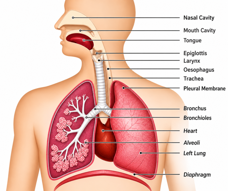
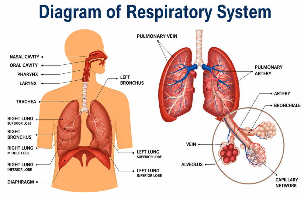
The main structures include:
- Nose and nasal cavity
- Mouth
- Pharynx (throat)
- Larynx (voice box)
- Epiglottis
- Trachea (windpipe)
- Bronchi
- Bronchioles
- Lungs
- Alveoli
- Diaphragm
Although its primary purpose is respiration, the respiratory system also helps us speak, smell and protect the body from harmful particles and microorganisms.
Understanding these structures becomes increasingly important as first aid training progresses. For example, learners completing our FREC 3 course develop their understanding of airway and respiratory assessment as part of a structured approach to casualty care.
The Journey of Air Through the Body
When you breathe in, air normally enters through the nose or mouth.
From there it travels through the:
Nose or mouth → pharynx → larynx → trachea → bronchi → bronchioles → alveoli
Each stage has an important role.
The nose helps warm, humidify and filter inhaled air before it travels deeper into the respiratory system.
Air then passes through the pharynx and larynx before entering the trachea.
At the bottom of the trachea, the airway divides into two main bronchi. One bronchus enters the right lung and the other enters the left.
These divide repeatedly into progressively smaller airways called bronchioles.
Eventually, the bronchioles terminate at tiny air sacs called alveoli.
It is here that one of the most important processes in the respiratory system takes place: gas exchange.
The Upper and Lower Airway
It can be useful to think of the respiratory tract as two main areas: the upper airway and lower airway.
The upper airway includes structures such as the nose, mouth, pharynx and larynx.
The lower airway includes the trachea, bronchi, bronchioles and structures within the lungs.
For a first aider, maintaining a clear airway is critical. An unconscious casualty, for example, may lose muscle tone and become unable to maintain their own airway effectively.
Airway assessment and management therefore form an important part of the primary survey, from basic first aid courses through to more advanced training such as FREC 3.
The Epiglottis
The respiratory and digestive systems share part of the same pathway, so the body needs a mechanism to help prevent food and fluid from entering the airway.
The epiglottis is a flap of cartilage situated near the entrance to the larynx.
During swallowing, it helps protect the airway and direct food and fluid towards the oesophagus rather than the trachea.
If a foreign object enters or blocks the airway, however, the casualty may begin choking.
Recognising and managing choking is therefore an essential practical skill taught during our Emergency First Aid at Work courses and other first aid qualifications.
The Trachea
The trachea, commonly called the windpipe, carries air towards the lungs.
It is supported by rings of cartilage that help prevent the airway from collapsing.
The inside of the respiratory tract also contains mucus and tiny hair-like structures called cilia. These form part of the body’s defence system.
Particles and microorganisms can become trapped in mucus, while the cilia help move the mucus upwards so that it can eventually be coughed out or swallowed.
The Bronchi and Bronchioles
Towards the bottom of the chest, the trachea divides into the right and left main bronchi.
Each bronchus enters a lung before dividing into progressively smaller branches.
These eventually become the bronchioles.
You can think of this structure rather like an upside-down tree:
Trachea → Bronchi → Bronchioles → Alveoli
The branching system allows air to be distributed throughout the lungs.
Conditions affecting these airways can make breathing considerably more difficult. In asthma, for example, narrowing and inflammation of the airways can restrict airflow and produce symptoms such as wheezing and breathlessness.
Recognising respiratory emergencies such as asthma forms part of both workplace and Outdoor First Aid training, where responders may also need to consider the consequences of being further away from emergency medical assistance.
The Lungs
The lungs are the main organs of the respiratory system and are located within the chest cavity.
The right lung has three lobes, while the left lung has two lobes. The left lung is slightly smaller because space within the chest is also occupied by the heart.
The lungs are surrounded by membranes called the pleura. Between these layers is a small amount of fluid that helps reduce friction as the lungs move during breathing.
The lungs themselves contain the extensive network of bronchioles and alveoli required for gas exchange.
What Are Alveoli?
At the end of the smallest bronchioles are microscopic air sacs known as alveoli.
These structures are extremely important because this is where oxygen and carbon dioxide are exchanged between the lungs and the bloodstream.
The alveoli have very thin walls and are surrounded by tiny blood vessels called capillaries.
When we inhale, oxygen reaches the alveoli and passes across the thin membrane into the blood.
At the same time, carbon dioxide moves in the opposite direction—from the blood into the alveoli—ready to be breathed out.
This process is known as gas exchange.
Why Does the Body Need Oxygen?
Oxygen is transported around the body in the blood, predominantly attached to haemoglobin within red blood cells.
The cardiovascular system then delivers this oxygenated blood to the body’s tissues.
Cells use oxygen as part of the process of producing the energy required for normal function.
This means the respiratory and cardiovascular systems are closely connected.
Simply getting air into the lungs isn’t enough. Oxygen must successfully cross into the bloodstream and then be circulated around the body.
This relationship becomes particularly important when learning about shock, major bleeding, respiratory emergencies and cardiac arrest.
These subjects are developed further during more advanced pre-hospital training such as our FREC 3 course, where learners build on basic first aid knowledge and develop a more systematic approach to patient assessment.
The Role of Carbon Dioxide
Carbon dioxide is produced as a waste product of cellular metabolism.
It is transported in the blood back towards the lungs, where it crosses into the alveoli and is removed when we breathe out.
Maintaining appropriate levels of carbon dioxide is important because carbon dioxide also contributes to the regulation of the body’s acid-base balance.
This is one reason changes in a patient’s respiratory rate or breathing pattern can provide important information about their condition.
The Diaphragm and Breathing
The lungs don’t inflate themselves.
Breathing relies heavily on the diaphragm, a large dome-shaped muscle situated underneath the lungs.
During inhalation
The diaphragm contracts and moves downwards.
This increases the volume within the chest and creates a pressure difference that draws air into the lungs.
During exhalation
The diaphragm relaxes and moves upwards.
The volume within the chest decreases and air moves out of the lungs.
Other muscles around the chest can also assist with breathing, particularly when the body’s demand for oxygen increases or when breathing becomes difficult.
A casualty who is working particularly hard to breathe may begin using these accessory muscles, which can be an important clinical observation.
Normal Breathing and Respiratory Assessment
For first aiders and pre-hospital responders, assessing breathing involves much more than simply deciding whether someone is breathing.
Depending on the responder’s level of training, assessment may include observing:
- Respiratory rate
- Depth of breathing
- Rhythm
- Chest movement
- Abnormal breathing sounds
- Ability to speak
- Skin colour
- Signs of increased respiratory effort
- Use of accessory muscles
- Oxygen saturation where appropriate equipment and training are available
A significant change in respiratory rate can sometimes be an early indication that a casualty’s condition is deteriorating.
This is why structured casualty assessment is an important part of our First Aid at Work training, and why learners progressing into pre-hospital care develop these assessment skills further.
What Happens When Breathing Fails?
If breathing becomes ineffective, the body may not receive enough oxygen.
There are many possible causes, including:
- Airway obstruction
- Choking
- Asthma
- Anaphylaxis
- Chest injury
- Pneumonia and other respiratory infections
- Drug overdose
- Head injury
- Reduced level of consciousness
- Cardiac arrest
Some conditions primarily affect the airway, while others affect ventilation, gas exchange, circulation or the body’s ability to use oxygen effectively.
Recognising these differences becomes increasingly important as a responder progresses beyond basic first aid.
Why Is the Respiratory System Important in First Aid?
Airway and breathing problems can become life-threatening very quickly.
During a primary survey, the casualty’s airway and breathing are assessed early so that immediately life-threatening problems can be identified and managed.
Someone completing a one-day Emergency First Aid at Work course needs to be able to recognise normal and abnormal breathing, manage an unconscious casualty, perform CPR and respond to choking.
Those undertaking First Aid at Work develop this knowledge further across a wider range of illnesses and injuries.
For people working in remote or outdoor environments, our 16-hour Outdoor First Aid course considers the additional challenges created when professional medical assistance may be some distance away.
At FREC 3 level, learners begin developing a much more detailed understanding of patient assessment, physiology and the management of medical and trauma emergencies.
The underlying principle, however, remains the same:
Without an effective airway and effective breathing, a casualty can deteriorate rapidly.
Key Points to Remember
The respiratory system brings oxygen into the body and removes carbon dioxide.
Air travels through the nose or mouth, pharynx, larynx and trachea before entering the bronchi and progressively smaller bronchioles.
At the end of the bronchioles are the alveoli, where oxygen passes into the bloodstream and carbon dioxide passes out.
The diaphragm is the primary muscle responsible for breathing.
The respiratory and cardiovascular systems work closely together to deliver oxygen to the body’s tissues.
For first aiders and pre-hospital responders, recognising changes in airway and breathing can provide vital early warning that a casualty is becoming seriously unwell.
Continue Learning
If you’re using the Optimal First Aid Knowledge Hub as FREC 3 pre-course reading, useful topics to explore next include the cardiovascular system, gas exchange, airway management, respiratory assessment, the primary survey (ABCDE) and shock.
These foundation topics will make the practical assessment and clinical content covered during your FREC 3 training considerably easier to understand once you move into the classroom.